The sensors capturing X-rays in hospitals today are the same technology recording your weekend portraits tomorrow. Medical imaging doesn’t just save lives in operating rooms—it revolutionizes the camera in your hands, driving innovations that trickle down to consumer photography within 18-24 months.
Consider how computed tomography sensors, designed to detect minute variations in tissue density, evolved into the back-illuminated CMOS sensors now standard in mirrorless cameras. These medical-grade components, originally engineered to process thousands of diagnostic images with zero margin for error, brought us the low-light capabilities that let you shoot concerts without grain and the dynamic range that preserves both shadow detail and highlight information in a single frame.
The pipeline works like this: researchers develop imaging solutions for detecting tumors at microscopic levels, requiring extreme sensitivity and precision. Defense contractors refine these technologies for satellite and surveillance applications. Finally, consumer electronics manufacturers adapt the proven designs, making them affordable for photography equipment. What costs $500,000 in a radiology suite becomes a $2,000 camera sensor within two product cycles.
Right now, medical facilities are deploying photon-counting detectors, AI-powered image reconstruction algorithms, and computational imaging systems that capture multiple exposures simultaneously. Each advancement addresses specific diagnostic challenges, but the underlying technology—the physics of light capture, signal processing, and noise reduction—translates directly to photography. Understanding these developments helps you anticipate which camera features will define the next generation of imaging tools.
The Tech Transfer Pipeline: From Hospital to Camera Bag
Why Medical Imaging Pushes Boundaries First
Medical imaging operates in a unique environment where innovation accelerates at remarkable speeds, and there’s a fascinating reason why. When lives hang in the balance, budgets expand dramatically. A hospital investing in diagnostic equipment isn’t shopping for the best price-to-performance ratio like a photographer choosing a new camera body. They’re acquiring technology that can detect a tumor at its earliest stage or reveal internal injuries that would otherwise remain hidden until it’s too late.
This life-or-death urgency creates what engineers call a “premium development environment.” Research teams receive funding that consumer electronics manufacturers can only dream about. A single MRI machine costs between $1 million and $3 million, and hospitals replace them every 7-10 years. That financial ecosystem supports aggressive R&D cycles.
Regulatory requirements also push innovation forward in unexpected ways. When the FDA demands that imaging sensors perform flawlessly across millions of diagnostic scans, manufacturers develop quality control processes and precision engineering techniques that eventually trickle down to consumer products. The same sensor technology that helps radiologists identify microscopic abnormalities in tissue becomes the foundation for your camera’s ability to capture clean images at ISO 12,800. Medical imaging doesn’t just push boundaries first; it establishes entirely new territories that photography equipment later explores with refinement and cost optimization.
Recent Crossover Success Stories
You’re probably already enjoying the benefits of medical imaging technology every time you press the shutter button. Let’s look at three breakthrough technologies that made the leap from hospital to camera bag.
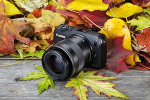
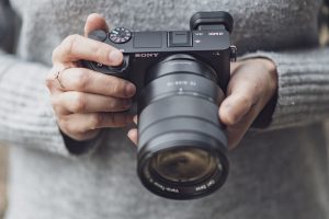
Backside-illuminated (BSI) sensors represent one of the most significant crossovers. Originally developed for X-ray imaging to capture more photons in low-radiation environments, BSI technology flips the traditional sensor design by moving the circuitry behind the photodiodes. This seemingly simple change dramatically improves light-gathering efficiency. Sony’s A7S series and many modern smartphones now use BSI sensors, giving photographers up to two stops better low-light performance compared to traditional sensors. That clean ISO 12,800 shot you captured at a dimly lit wedding? Thank medical imaging research.
Phase detection autofocus systems also originated in medical imaging, where doctors needed to rapidly focus on specific tissue layers during endoscopic procedures. Camera manufacturers adapted these same principles to create on-sensor phase detection points. Today’s mirrorless cameras can have hundreds of AF points covering nearly the entire frame, tracking subjects with precision that seemed impossible just a decade ago.
Perhaps most impressive is computational noise reduction. Medical scanners needed to reduce radiation exposure while maintaining image quality, leading to sophisticated denoising algorithms. These same mathematical models now power the noise reduction in your camera and editing software. Modern cameras apply multi-frame processing borrowed directly from CT scan technology, stacking multiple exposures in milliseconds to produce remarkably clean high-ISO images that would have been unusable just five years ago.
What’s Breaking Through Right Now: Latest Medical Imaging Innovations
Photon-Counting Detectors: The Next Sensor Revolution
Imagine a sensor so sensitive it can count individual photons – literally particles of light – as they arrive. That’s exactly what photon-counting detectors (PCDs) do in the latest medical CT scanners, and this technology represents a fascinating glimpse into photography’s potential future.
Traditional medical CT scanners, like conventional camera sensors, integrate light energy over a period. They measure the total amount of radiation that hits the detector but can’t distinguish individual photons. Photon-counting detectors flip this approach entirely. By registering each photon separately and measuring its energy level, these sensors deliver unprecedented image quality with dramatically reduced radiation doses – sometimes up to 40% less than conventional systems.
Here’s where it gets exciting for photographers: the core principles solving medical imaging challenges mirror problems we face daily. Low-light photography struggles with noise because sensors can’t efficiently separate signal from random electronic interference. Photon-counting technology essentially eliminates this noise floor by only registering actual light particles, not electrical artifacts.
The dynamic range implications are particularly compelling. Since PCDs can measure each photon’s energy, they theoretically capture a much wider tonal range in a single exposure – imagine pulling detail from deep shadows and bright highlights without the compromises of current sensors. Companies like Canon and Sony are already researching similar photon-counting approaches for consumer cameras, though mainstream availability remains years away.
While we’re not quite ready to buy photon-counting cameras, understanding this technology helps contextualize where sensor development is headed: toward fundamentally different approaches that count light rather than simply measuring it.
AI-Powered Image Reconstruction
One of the most exciting crossovers from medical imaging to photography is AI-powered image reconstruction. In hospitals, radiologists face a constant challenge: getting diagnostic-quality images while minimizing patient exposure to radiation or lengthy scan times. The solution? Artificial intelligence that can essentially fill in the gaps, creating crystal-clear images from incomplete data.
Here’s where it gets interesting for photographers. The same AI algorithms used in MRI and CT scanners are now making their way into computational photography. Think about it: when you shoot in extreme low light and your camera produces a surprisingly clean image, you’re benefiting from similar technology. These AI systems learn to recognize patterns and textures, then intelligently reconstruct detail that would otherwise be lost to noise or underexposure.
Medical imaging’s real-time enhancement capabilities are particularly promising. Surgeons now use AI-enhanced imaging during procedures, getting instant feedback with improved clarity. This same concept is appearing in mirrorless cameras with real-time denoising and in smartphone photography apps that process images on the fly.
The practical implication? Your next camera upgrade will likely include neural processing units specifically designed for image reconstruction. We’re moving beyond traditional noise reduction into territory where AI can actually recover detail from seemingly impossible shooting conditions, all because medical researchers needed to see tumors more clearly with less radiation exposure.
Advanced Multispectral Imaging
Medical imaging routinely captures wavelengths invisible to our eyes, and this technology is now trickling down to consumer cameras. Infrared imaging, for instance, helps surgeons visualize blood flow during procedures by detecting heat signatures. Similarly, hyperspectral cameras used in dermatology can identify skin cancers by analyzing how tissues reflect different wavelengths of light. These same principles are appearing in mainstream photography through enhanced sensor designs.
Modern camera sensors increasingly incorporate extended spectral sensitivity, borrowing from medical technology that needed to see beyond RGB. Some mirrorless cameras now offer improved infrared response for specialized photography, while computational imaging combines multiple wavelength data to reveal details traditional sensors miss. Medical imaging’s push for better low-light performance in operating rooms has directly influenced backside-illuminated sensor development you’ll find in today’s cameras.
The practical takeaway? Those remarkable advances in smartphone night mode and astrophotography didn’t emerge in isolation. They’re built on decades of medical research demanding sensors that capture more information across broader spectral ranges, now accessible to everyday photographers.
The Regulatory Landscape: Why It Matters to You
How Medical Device Approval Accelerates Consumer Tech
When a medical device receives regulatory approval from agencies like the FDA or CE marking in Europe, it’s essentially earned a gold star for safety and effectiveness. This matters tremendously for photography because it creates a proven pathway for technology to migrate into consumer cameras.
Think of regulatory validation as rigorous beta testing at an extreme level. Medical imaging sensors must perform flawlessly in life-or-death situations, enduring millions of exposures while maintaining color accuracy and dynamic range. When these sensors pass clinical trials, camera manufacturers gain confidence that the underlying technology is mature, reliable, and safe for mass production.
The regulatory process also establishes manufacturing standards and quality control protocols that directly benefit consumer electronics. A sensor approved for medical use comes with documented performance metrics, safety data, and production consistency requirements. Camera makers can leverage this groundwork rather than starting from scratch, significantly reducing their research and development costs and time to market.
Perhaps most importantly, regulatory approval proves real-world viability beyond laboratory conditions. Medical devices operate in demanding environments with zero tolerance for failure, so their certification demonstrates that technologies like advanced CMOS sensors, sophisticated noise reduction algorithms, and AI-powered image processing aren’t just conceptually sound but practically deployable. This validation removes much of the risk manufacturers face when deciding whether to invest in new imaging technologies for their next camera generation.
Recent Policy Shifts Opening New Doors
The regulatory landscape has shifted dramatically over the past year, and if you’re wondering why that matters to photographers, here’s the exciting part: medical imaging regulations often pave the way for what eventually lands in your camera bag.
In early 2024, the FDA finalized its comprehensive framework for AI and machine learning in medical imaging systems. This wasn’t just bureaucratic paperwork—it created a fast-track approval pathway that’s already bearing fruit. The guidelines established clear standards for adaptive algorithms that learn and improve over time, similar to how computational photography in modern cameras continuously refines image processing. Several AI-powered diagnostic imaging systems received approval within months rather than years, demonstrating how streamlined oversight can accelerate innovation without compromising safety.
The European Union followed suit in late 2024 with updated Medical Device Regulation amendments specifically addressing photon-counting CT scanners and other next-generation modalities. These aren’t incremental improvements—they represent fundamental shifts in how we capture light information, much like the jump from CCD to CMOS sensors revolutionized consumer photography.
Perhaps most relevant to photographers, the approval process for new sensor technologies has been expedited. Companies developing advanced silicon photomultipliers and quantum dot sensors for medical applications can now navigate regulatory hurdles more efficiently. The practical outcome? Technologies that would have languished in medical-only applications for a decade are reaching consumer imaging markets within three to five years. We’re already seeing trickle-down effects in smartphone cameras and mirrorless systems, with several manufacturers announcing sensor technologies that originated in these recently approved medical imaging platforms.
What This Means for Your Photography Gear (The Practical Outlook)

Technologies to Watch in the Next Camera Release Cycle
Based on recent FDA approvals and medical imaging breakthroughs, several exciting features should reach consumer cameras by 2025-2026. Here’s what to watch for on your next upgrade cycle.
Quantum dot sensor technology, recently approved for mammography systems, promises significantly improved dynamic range. Early prototypes show a potential 2-3 stop improvement in shadow detail recovery without the noise penalty we’ve seen in traditional silicon sensors. Expect this in flagship models first, then trickling down to mid-range bodies within 18 months.
AI-powered noise reduction is another crossover worth monitoring. Medical CT scanners now use machine learning algorithms that can distinguish between diagnostic information and noise in real-time. Camera manufacturers are already testing similar systems that could deliver usable ISO 102,400 images without the “plastic” look of current high-ISO shots. This isn’t your standard computational photography trick—it’s genuine signal recovery based on pattern recognition trained on billions of image samples.
Time-of-flight autofocus represents the most tangible near-term upgrade. Originally developed for surgical navigation systems, this technology measures actual distance using light pulses rather than contrast detection. Translation for photographers: instant focus acquisition in complete darkness and tracking accuracy that doesn’t falter with low-contrast subjects like black cats or wedding dresses.
Phase-detection pixels with 100 percent sensor coverage are moving from prototype to production, borrowed directly from retinal imaging systems. Combined with the processing advances mentioned above, we’re looking at a genuine leap forward rather than incremental improvements.
Should You Wait or Buy Now?
The million-dollar question: should you hold off on that camera purchase, or pull the trigger now? The answer depends entirely on where you sit in the photography spectrum.
If you’re a professional relying on your gear for income, waiting isn’t realistic. The imaging innovations we’ve discussed are already filtering into current models, and the cameras available today represent a massive leap from just five years ago. You need equipment that works now, not hypothetical sensors arriving in two years. Plus, the technology adoption curve means truly groundbreaking advances take 3-5 years to become affordable and reliable enough for professional use.
For enthusiast photographers with recent gear (purchased within the last 2-3 years), there’s little urgency. Your camera likely already benefits from early-generation versions of these medical imaging adaptations. The improvements coming in the next product cycle will be incremental rather than revolutionary. Focus on mastering what you have and investing in glass, which holds value longer than bodies.
However, if you’re working with older equipment (5+ years) and considering an upgrade anyway, timing your purchase strategically makes sense. Major manufacturers typically announce flagship models on predictable cycles. Waiting 3-6 months for the next generation could net you significantly better low-light performance and computational photography features derived from these medical imaging breakthroughs.
Beginning photographers should buy now and buy smart. Entry-level and mid-range cameras already incorporate many sensor advances we’ve discussed. The learning curve matters more than having cutting-edge technology, and today’s “good enough” far exceeds yesterday’s professional standards.
The reality is that there will always be something better on the horizon. The best camera remains the one you actually have and use, not the one you’re waiting to buy.
The connection between medical imaging and consumer photography isn’t just interesting trivia—it’s a window into your camera’s future. As we’ve explored, technologies like advanced CMOS sensors, computational photography algorithms, and AI-powered image processing all follow a remarkably consistent path from hospital to handheld device. Understanding this pipeline gives you a genuine advantage when evaluating new gear, anticipating market trends, and making informed purchasing decisions.
Here’s your practical takeaway: start following the FDA’s medical device approvals and the EC’s medical device regulations, particularly announcements related to imaging sensors and AI diagnostic tools. When you see terms like “back-illuminated sensors,” “photon-counting detectors,” or “deep learning image reconstruction” in medical imaging news, bookmark those stories. Within 18 to 36 months, you’ll likely encounter similar technology in camera announcements, often with the same underlying science adapted for creative applications.
The convergence we’re witnessing is genuinely exciting. Medical imaging researchers are solving the same fundamental problems photographers face—capturing clean images in challenging conditions, extracting maximum detail from limited light, and processing vast amounts of data intelligently. Their solutions, proven in demanding clinical environments, eventually become the features that transform how you shoot.
Stay curious about developments beyond photography’s traditional boundaries. The next breakthrough in low-light performance or autofocus accuracy might be making headlines in a radiology journal right now, waiting for someone attentive enough to recognize its potential. That someone could be you.