 By
By 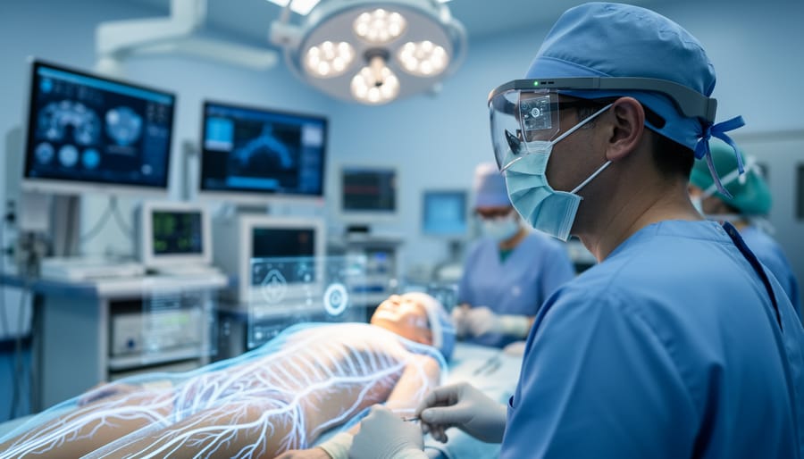
Augmented reality in healthcare is a technology that overlays digital information, three-dimensional models, or real-time data onto a clinician’s view of the physical world, typically through specialized glasses, headsets, or camera-enabled devices. Instead of replacing what a surgeon or nurse sees, AR enhances it by adding layers of critical information exactly where and when it’s needed.
This isn’t science fiction playing out in research labs anymore. In 2026, AR systems guide surgeons through complex procedures by projecting vein maps and tumor boundaries directly onto a patient’s body. Medical students practice anatomy on virtual cadavers that appear to float above their desks. Emergency responders receive step-by-step resuscitation instructions overlaid on actual patients. The technology has moved from experimental trials to operating rooms, ambulances, and clinics around the world.
What makes this possible is the same core imaging technology that photographers have relied on for years. AR camera technology uses advanced sensors, depth-mapping cameras, and processing algorithms to understand three-dimensional space in real time. These systems track movement, recognize anatomical structures, and anchor digital overlays to physical objects with millimeter precision. The difference between a powerful diagnostic tool and a gimmick often comes down to camera quality, sensor accuracy, and processing speed.
Understanding how AR actually functions in medical settings requires looking beyond the flashy headlines. The camera systems capture the real world, specialized software interprets what those cameras see, and display technology presents the augmented view back to the user. Each component plays a role in determining whether AR becomes a practical tool or an expensive distraction. This article breaks down the mechanics, explores the different categories of medical AR applications, and examines real examples of how hospitals and clinics are putting this technology to work right now.
What Augmented Reality Means in Medical Settings
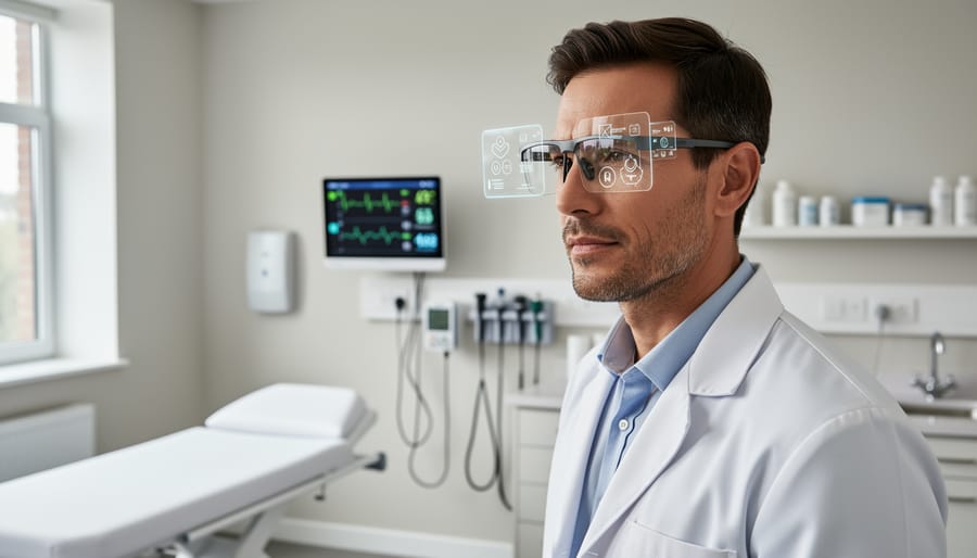
Augmented reality in healthcare works by adding computer-generated images, data, or guidance directly on top of what a doctor or medical professional sees in the real world. Unlike virtual reality, which places users in a completely artificial environment, AR enhances the existing physical space by superimposing useful information where it’s needed most, on a patient’s body during surgery, over medical equipment during training, or alongside a practitioner’s field of view during diagnosis. Augmented versus immersive systems differ fundamentally in that AR keeps clinicians grounded in their actual environment while adding digital layers that would otherwise be invisible.
The technology relies on cameras and sensors to understand the physical space and track objects within it. A typical medical AR system uses one or more cameras to capture the real-world scene, whether that’s an operating table, a patient’s arm, or a training model. Depth sensors measure distances and create three-dimensional maps of surfaces, while tracking algorithms continuously update the position and orientation of both the user and the objects being augmented. This spatial awareness allows the system to place digital overlays in precisely the right location and keep them anchored there as people and cameras move.
- Augmented Reality
- Technology that overlays digital information onto the real world, visible through displays like headsets, tablets, or specialized glasses.
- Medical Imaging
- The process of creating visual representations of internal body structures, such as CT scans or MRI data, which AR can overlay onto a patient’s physical anatomy.
- Spatial Mapping
- The technique cameras and sensors use to build a three-dimensional understanding of the physical environment, enabling accurate placement of digital overlays.
- Depth Sensing
- Camera technology that measures the distance to objects in a scene, creating 3D maps that help AR systems understand surfaces and contours.
- Real-time Overlay
- The continuous rendering and repositioning of digital graphics so they remain aligned with physical objects as the user or environment moves.
What makes AR particularly valuable in medicine is its ability to show clinicians information they couldn’t otherwise see without looking away from the patient or switching between screens. Instead of glancing at a monitor across the room to check a CT scan during surgery, a surgeon can see that same scan projected directly onto the patient’s body. The camera system tracks the patient’s position, and the software adjusts the overlay in real time, maintaining alignment even as the surgical team repositions instruments or adjusts lighting. This seamless integration of digital data with physical reality creates a new way of working that keeps attention focused where it matters most.
How AR Camera Systems Work in Healthcare
Camera and Sensor Requirements
Medical-grade AR systems demand camera hardware far beyond what consumer devices offer. The core requirements start with high-resolution RGB cameras, typically 4K or better, that capture clear anatomical detail even under variable surgical lighting. These cameras must maintain accurate color reproduction, since tissue differentiation often depends on subtle hue variations that guide surgical decisions.
Equally critical are depth-sensing cameras that create three-dimensional maps of the operating field. Time-of-flight sensors and structured light systems measure distances with sub-millimeter precision, enabling AR overlays to align correctly with patient anatomy as the surgeon’s viewpoint shifts. This spatial accuracy requires depth cameras capable of rapid refresh rates, often 60Hz or higher, to track movement without lag.
Infrared tracking cameras add another layer, using active or passive markers to monitor instrument positions and head movement in real-time. Multi-camera arrays positioned around the surgical field provide overlapping coverage, eliminating blind spots and maintaining tracking even when one camera’s view is temporarily obscured by hands or instruments.
Medical AR headsets typically integrate four to six cameras working simultaneously: front-facing RGB for scene capture, outward-facing depth sensors for spatial mapping, downward-angled cameras for hand tracking, and sometimes dedicated eye-tracking cameras to detect where the surgeon is looking. This multi-sensor fusion creates the robust spatial awareness healthcare applications require, processing data from all cameras to build a coherent, stable augmented view.
Image Processing and Registration
Once AR cameras capture depth and visual data from a patient’s body, sophisticated algorithms transform that raw input into precisely aligned digital overlays. This processing pipeline typically unfolds in three critical stages.
First comes calibration, the system establishes baseline reference points between the camera’s view and the patient’s anatomy. Medical AR platforms use distinctive anatomical landmarks or external markers placed on the patient’s skin to create this initial spatial map. The software calculates exactly how the camera sits relative to the body, accounting for lens distortion and perspective.
Tracking algorithms then maintain this registration as the camera or patient moves. Most systems employ simultaneous localization and mapping (SLAM) techniques borrowed from robotics, continuously updating the spatial relationship by identifying features in the camera feed. High-end surgical AR uses infrared markers or bone-mounted reference frames that cameras track at 60+ frames per second, ensuring overlays don’t drift during procedures.
The rendering pipeline completes the process by generating the digital overlay and positioning it frame by frame. Processing units, often built into AR headsets or connected workstations, blend CT scan slices, blood vessel maps, or anatomical models with the live camera view. Depth data ensures virtual elements appear behind or in front of real structures appropriately.
Latency matters enormously here. Delays beyond 20 milliseconds between camera input and rendered output create misalignment that surgeons can’t trust, so medical AR systems prioritize real-time processing over visual complexity.
Categories of AR Camera Applications in Medicine
Surgical Navigation and Visualization
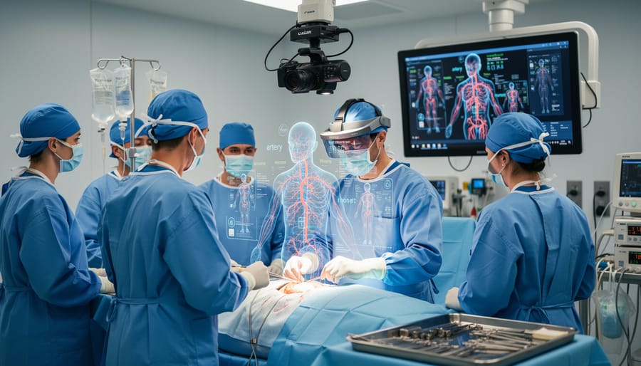
Surgical AR represents one of the most technically demanding applications of camera-based augmented reality in medicine. During complex procedures, neurosurgery, orthopedic reconstruction, tumor resection, surgeons use AR headsets or mounted displays that project preoperative scan data directly onto the patient’s body. The system relies on advanced computer vision tech to track both the patient’s position and the surgeon’s viewpoint in real time, ensuring that the digital overlay of internal structures stays precisely aligned as people move around the operating table.
AR navigation using CT/MRI scans lets surgeons see beneath the surface without breaking eye contact with the patient, no need to glance at a separate monitor across the room. Multi-camera arrays mounted on the headset or surgical microscope track infrared markers placed on the patient or instruments, recalculating registration hundreds of times per second. This continuous tracking compensates for breathing motion, small shifts in patient position, and the surgeon’s own movement, maintaining millimeter-level accuracy throughout the procedure.
Vein and Vasculature Imaging
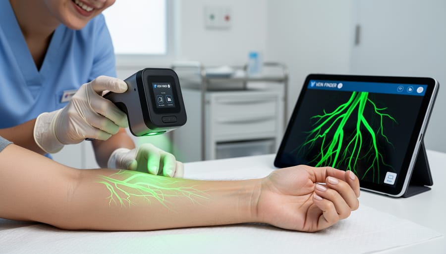
Near-infrared camera systems have revolutionized one of healthcare’s most common procedures: finding veins for IV insertion and blood draws. These AR devices use specialized cameras that detect hemoglobin’s light absorption properties, making subcutaneous blood vessels visible up to 10mm beneath the skin surface, far deeper than the naked eye can perceive.
The camera captures near-infrared light reflected from tissue, processes the image to identify vascular patterns, and projects a real-time map of veins directly onto the patient’s skin using visible light. This overlay moves with the patient, maintaining accurate registration as clinicians adjust positioning.
The technology proves particularly valuable for patients with difficult venous access, those with dark skin tones, obesity, chronic illness, or previous chemotherapy where traditional visual and palpation methods often fail. Emergency departments and pediatric units report first-stick success rates improving from 60% to over 85% with vein visualization systems.
Devices like AccuVein and VeinViewer use compact LED projectors synchronized with their infrared cameras, creating a portable system that works in various lighting conditions. The camera refresh rate matters here: faster frame rates maintain overlay accuracy during movement, preventing misalignment between the projected image and actual vein location.
Medical Education and Training
Medical schools and training programs now use AR camera systems to layer realistic 3D anatomy onto physical spaces, letting students examine organs and systems without a cadaver. A tablet or AR headset tracks the position of a training mannequin or even a real patient volunteer, then projects detailed anatomical structures, muscles, nerves, blood vessels, onto the surface. Students can walk around the model, zoom into specific areas, and peel back layers to see how structures relate spatially. This camera-based approach turns any classroom into an anatomy lab. Programs like Case Western Reserve University’s HoloAnatomy use depth-sensing cameras to anchor holographic models in space, while simpler tablet-based platforms rely on marker tracking to position overlays accurately.
Patient Rehabilitation and Physical Therapy
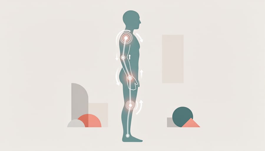
AR rehabilitation systems use depth-sensing cameras, similar to those in smartphone portrait modes but calibrated for full-body motion capture, to track patient movements during physical therapy exercises. These cameras measure joint angles, range of motion, and movement patterns in real time, comparing them against prescribed therapeutic targets. The system projects visual guides onto a screen or through headset displays, turning repetitive exercises into interactive games that respond to proper form.
A stroke patient relearning arm movement might see virtual objects to reach for, with the camera detecting whether their shoulder rotation and elbow extension match recovery goals. The immediate visual feedback helps patients self-correct without waiting for a therapist’s assessment. Many systems record motion data across sessions, letting clinicians track progress through precise metrics rather than subjective observation. This camera-driven approach addresses a core rehabilitation challenge: maintaining correct form during home exercises when supervision isn’t available.
Real-World Medical Applications of AR Camera Technology
Operating Room Enhancement
During complex procedures, surgeons traditionally had to shift their gaze between the patient and external monitors displaying CT or MRI scans. AR surgical systems eliminate this disruption by projecting three-dimensional anatomical data directly onto the surgical field.
Microsoft’s HoloLens 2 has become a foundation for several FDA-cleared surgical platforms. These headset-mounted systems use multiple depth cameras and spatial sensors to track the patient’s position in real time, overlaying tumor boundaries, vascular structures, or bone anatomy precisely where they exist beneath the skin. Spine surgeons, for instance, can see vertebrae placement and screw trajectories without looking away, reducing procedural time and improving accuracy.
Proprietary navigation systems from companies like Brainlab and Stryker employ ceiling-mounted or instrument-integrated cameras that create persistent 3D holograms in the surgeon’s field of view. These platforms continuously adjust the overlay as the patient or surgeon moves, maintaining millimeter-level precision throughout procedures.
The camera arrays in these systems typically combine RGB imaging with infrared tracking markers attached to instruments or patient anatomy, creating a stable reference frame even as surgical conditions change. This fusion of camera technologies transforms static preoperative scans into dynamic, interactive surgical guidance.
Emergency and Clinical Settings
In emergency departments and clinical settings where speed and accuracy are critical, portable AR devices have become valuable tools for procedures that traditionally required multiple attempts or specialized expertise. Vein finder systems use near-infrared cameras operating at wavelengths between 740-850nm to detect hemoglobin beneath the skin, then project a real-time map of veins directly onto the patient’s arm or hand. These devices work across diverse lighting conditions, from bright fluorescent ER lights to dimly lit ambulances, because NIR sensors aren’t affected by ambient visible light the same way standard cameras are.
For ultrasound guidance, AR platforms like GE Healthcare’s Venue systems overlay ultrasound imagery onto the patient’s body using tablet cameras with depth sensors, helping clinicians visualize needle paths during central line placements or nerve blocks. The combination of wide dynamic range sensors and fast processing (typically 30fps minimum) ensures the overlay remains stable even when the clinician’s hands shake slightly during time-sensitive procedures.
Rapid triage assessment tools use smartphone or tablet cameras with AR software to measure vital signs, detect asymmetries, or guide trauma protocols, functioning reliably despite the chaotic, unpredictable conditions emergency teams face daily.
Remote Expertise and Telemedicine
AR cameras are reshaping how specialists collaborate across distances. When a rural clinic faces a complex case, an on-site provider can use a headset or tablet camera to stream their view to a specialist hundreds of miles away. The remote expert sees exactly what the local clinician sees, the patient, the wound, the scan images on a nearby monitor, and can draw arrows, highlight areas of concern, or place virtual markers directly into that live view.
These annotations appear in the on-site provider’s field of vision in real time, guiding their hands during a procedure or helping them identify anatomical landmarks. Systems like Proximie and Visionable use standard cameras enhanced with spatial tracking to anchor digital annotations to physical locations, so markings stay aligned even as the camera moves.
This approach dramatically reduces referral delays and travel burdens, particularly for underserved areas lacking specialist coverage. A cardiologist can guide catheter placement, a dermatologist can assess a suspicious lesion, or a trauma surgeon can direct emergency stabilization, all without leaving their office. The camera becomes a shared visual workspace where expertise flows instantly to wherever it’s needed most.
Camera Technology Behind Healthcare AR Systems
Medical AR systems rely on camera technology that goes far beyond what you’d find in even high-end consumer photography gear. The precision required to overlay digital information onto living tissue, often during life-or-death procedures, demands imaging capabilities that push the boundaries of what cameras can do.
At the hardware level, medical AR platforms typically combine multiple camera types in a single system. RGB cameras capture visible light for scene understanding and tracking, while depth sensors, often using structured light or time-of-flight technology, measure the precise distance to every point in the field of view. This depth data is what allows real-time overlays to maintain accurate positioning as a surgeon’s head moves or a patient breathes. Infrared cameras add another layer, tracking reflective markers placed on instruments or anatomy to maintain sub-millimeter registration accuracy.
The frame rates and latency requirements separate medical AR cameras from conventional imaging systems. A surgical navigation system might process camera feeds at 90-120 frames per second with end-to-end latency under 20 milliseconds, any lag between head movement and display update would cause nausea in the user or dangerous misalignment of projected anatomy. This demands not just fast sensors, but optimized image processing pipelines that prioritize speed without sacrificing the accuracy needed for medical decisions.
Camera calibration in these systems is also far more rigorous than standard photography applications. Medical AR platforms undergo multi-stage calibration processes that account for lens distortion, sensor alignment, and environmental factors like operating room lighting. Many systems include continuous self-calibration routines that adjust for thermal drift in the camera sensors, a phenomenon that could shift overlay accuracy by several millimeters over the course of a long surgery.
The computational photography techniques used in medical AR have started to influence consumer camera development. Multi-camera fusion algorithms that combine depth and color data, advanced tracking systems that maintain lock on moving subjects, and AI-enhanced image processing that extracts meaningful information from complex scenes, all of these innovations flow in both directions between medical imaging and the cameras photographers use every day.
Common Questions About AR in Healthcare
Healthcare professionals and institutions considering AR camera systems naturally have questions about their practical implementation. While the technology shows tremendous promise, understanding its current limitations and real-world constraints helps set realistic expectations.
How accurate are AR medical systems?
Modern medical AR systems achieve sub-millimeter accuracy when properly calibrated, with surgical navigation platforms typically maintaining 1-2mm precision throughout procedures. However, accuracy depends heavily on camera quality, calibration protocols, and stable tracking conditions, movement, lighting changes, or occlusions can temporarily degrade performance.
Are AR cameras safe for patients?
Yes, the camera systems themselves pose no direct safety risk, as they use standard visible light and near-infrared wavelengths already common in medical imaging. The primary safety considerations involve ensuring AR overlays don’t mislead clinicians and maintaining sterile fields when cameras are used in operating rooms.
What prevents wider adoption of AR in healthcare?
Cost remains the biggest barrier, as complete AR surgical systems can run $50,000-$500,000, followed by integration challenges with existing hospital IT systems and the learning curve for clinical staff. Regulatory approval processes also slow deployment of new AR applications.
Will smartphones replace specialized AR medical cameras?
Unlikely for critical applications, though smartphones are useful for education and some clinical tasks. Medical-grade AR systems require specialized depth sensors, infrared cameras, and processing power beyond current smartphone capabilities, plus they must meet stringent accuracy and reliability standards that consumer devices aren’t designed for.
The accuracy question particularly matters for surgical applications. While manufacturers advertise impressive precision figures, real-world performance varies with conditions. A surgeon moving quickly, variable operating room lighting, or blood on tracking markers can all affect how well the camera maintains registration between digital overlays and physical anatomy. Most systems include visual indicators when tracking confidence drops below acceptable thresholds, preventing clinicians from relying on potentially misaligned information.
Cost considerations extend beyond hardware. Training programs, software licensing, maintenance contracts, and IT integration can double the initial equipment investment. Smaller hospitals and clinics often start with lower-cost vein visualization cameras before committing to comprehensive surgical AR platforms.
The smartphone question reflects broader trends in medical technology. While consumer devices have driven AR adoption in other fields, healthcare’s margin for error is far smaller. A 5mm tracking error might be acceptable for furniture placement in a home design app but completely unacceptable when guiding a surgical instrument near vital structures. Dedicated medical AR cameras will likely remain necessary for high-stakes applications, even as smartphone-based systems find roles in training, patient education, and preliminary assessment tasks where extreme precision matters less than accessibility and ease of use.
Augmented reality in healthcare stands as one of the most compelling demonstrations of how camera and imaging technology can directly save lives and improve patient outcomes. The AR medical systems transforming surgery, diagnosis, and treatment today rely on the same fundamental principles that drive innovation in photography, precise sensor calibration, advanced image processing, accurate color reproduction, and real-time computational photography.
For anyone passionate about camera technology, the healthcare AR space offers a fascinating glimpse into where imaging innovation is headed. The multi-camera arrays tracking surgeon hand movements in operating rooms, the near-infrared sensors mapping vein networks beneath skin, and the depth-sensing systems registering digital overlays onto physical anatomy all represent cutting-edge applications of photographic principles you already understand.
What makes this convergence particularly exciting is how it pushes camera technology forward. The demands of medical AR, submillimeter accuracy, zero-latency processing, operation in challenging lighting conditions, and absolute reliability, drive sensor and lens development that eventually filters into consumer and professional photography equipment. The computational photography techniques pioneered for surgical navigation will likely influence how your next camera processes and displays information.
As AR headsets become lighter, camera sensors grow more sophisticated, and processing power continues advancing, the line between medical imaging and traditional photography will continue blurring. The skills and knowledge photographers develop understanding exposure, focus, sensor behavior, and optical physics translate directly to comprehending and potentially contributing to this emerging field.
Healthcare AR represents camera technology at its most consequential, where the precision of imaging systems directly impacts human health. That intersection makes it worth watching, regardless of whether you shoot portraits or surgical procedures.




 By
By